
Patient Information
Basical Information
Male, 55 years
Chief complaint
History of Present Illness
Past Medical History
Neurological Examination
Findings: Mixed aphasia, dysarthria, right facial droop, RUE strength 1/5, RLE 4−/5, right Babinski sign positive.
Prooperative Diagnosis
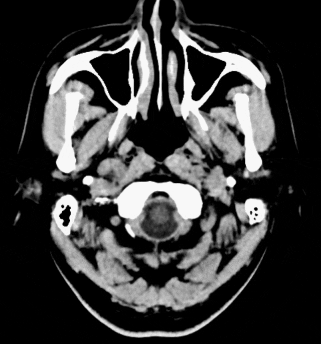
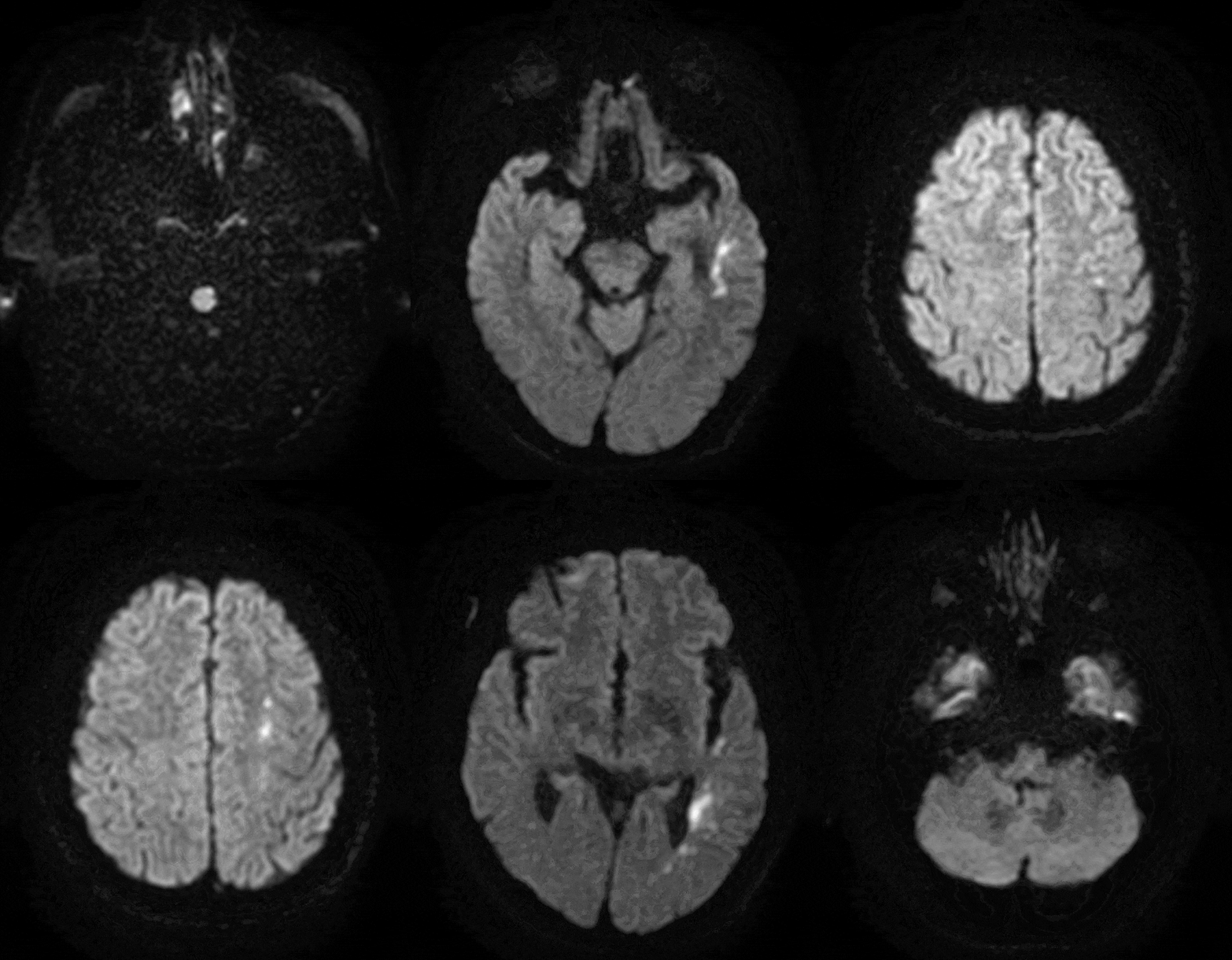
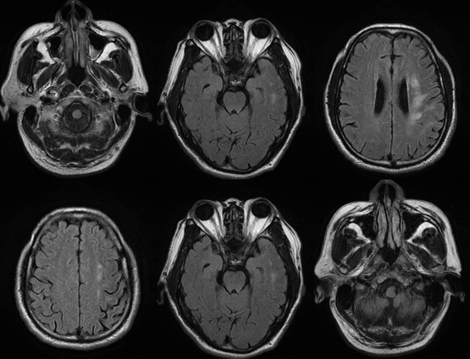
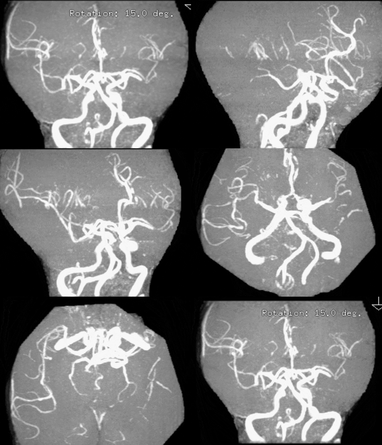
360° tortuous C1 segment of the left internal carotid artery
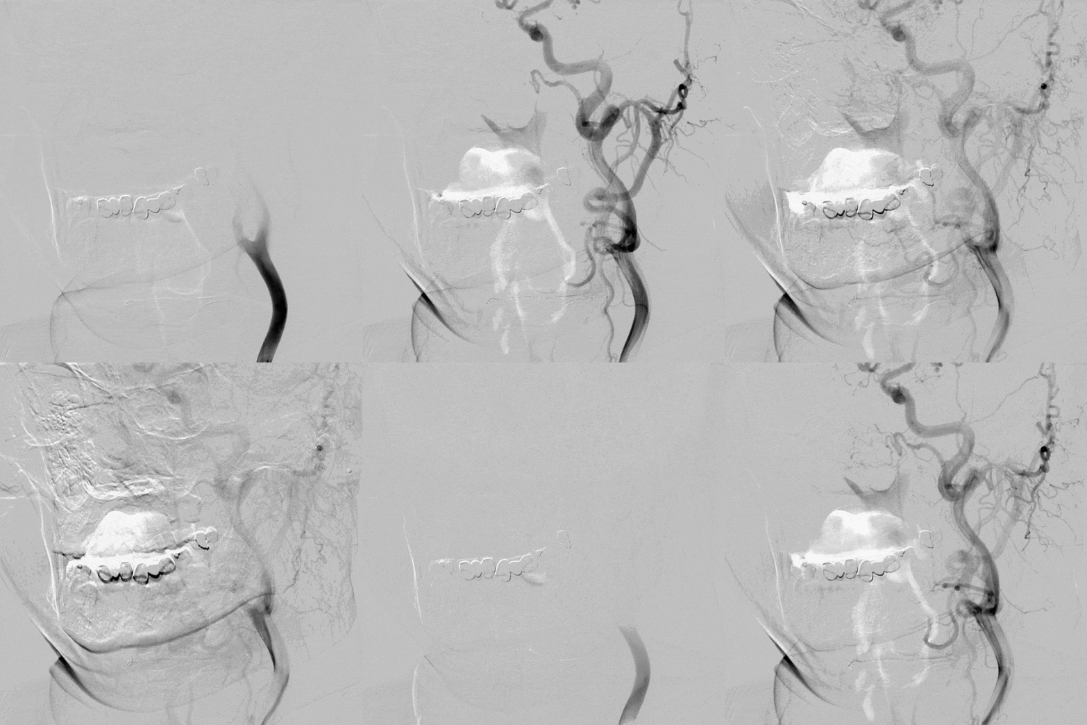
Left internal carotid artery access route
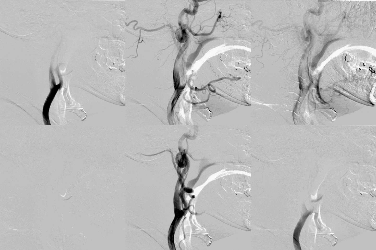
Left internal carotid artery access route
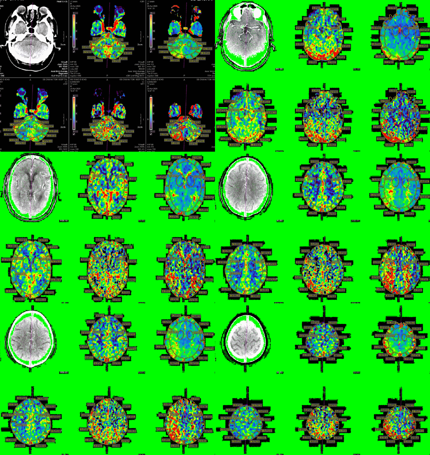
Severe stenosis of the M1 segment of the left middle cerebral artery with a second recurrence despite medical therapy
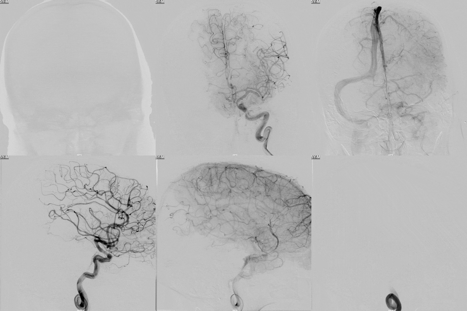
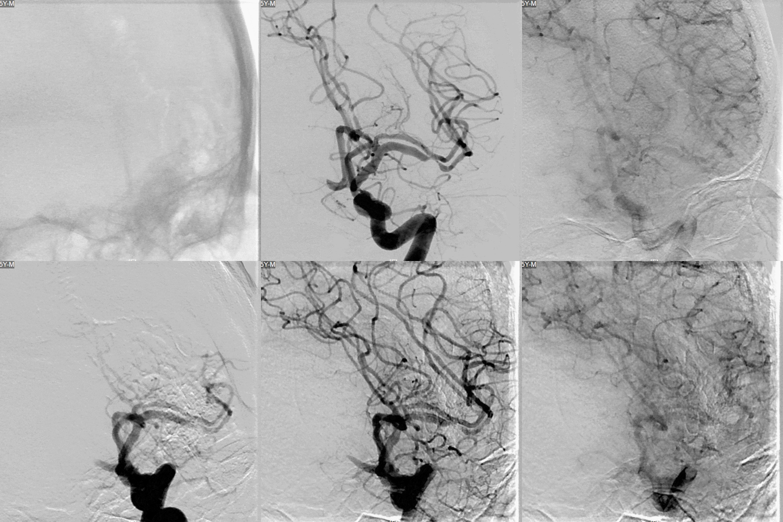
Diagnosis
Severe stenosis of the M1 segment of the left middle cerebral artery
Acute cerebral infarction
Grade 3 hypertension, very high-risk group
Type 2 diabetes mellitus
Hyperlipidemia
Surgery
Surgical Plan
Procedure:
Balloon angioplasty followed by stent placement in the M1 segment of the left middle cerebral artery under general anesthesia with endotracheal intubation.
Procedural Challenges:
The left internal carotid artery showed marked tortuosity with a Type III cavernous carotid configuration.
Preoperative Medication
- Cilostazol 100 mg bid
Clopidogrel 75 mg qd
Atorvastatin calcium 40 mg qn
Surgical Devices
- Locaste 8F Guiding Catheter, 100 cm
- Diagnostic Catheter, 125 cm
Radifocus Guidewire (hydrophilic guidewire)
6F Distal Access Catheter, 125 cm
Intracranial Balloon Dilatation Catheter, 2.0 × 12 mm
SL-10 Microcatheter
Guidewire 0.014“
Intracranial Drug-Eluting Stent, 2.5 × 12 mm
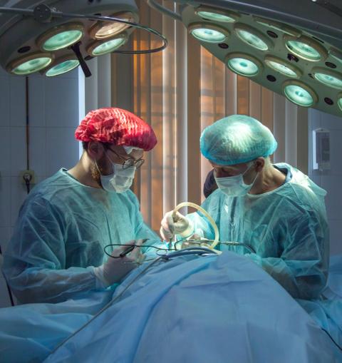
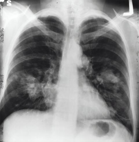
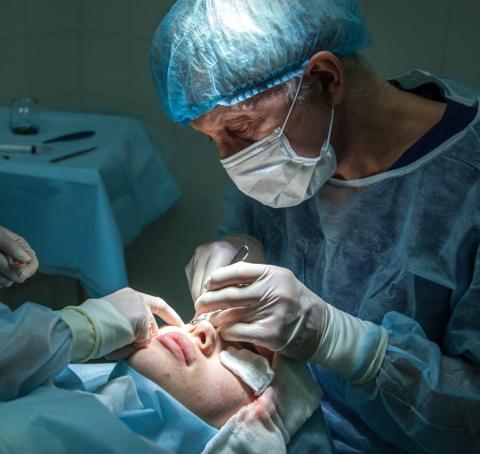
Operator Experience
Severe vascular tortuosity is a major challenge in endovascular treatment of intracranial arterial stenosis and, in extreme cases, may result in procedural failure. The use of more flexible distal access systems, such as 5F or 6F distal access catheters combined with a 6F long sheath, can facilitate navigation through tortuous vascular anatomy. However, careful consideration must be given to the working length of the system to ensure that the balloon or stent delivery device can adequately reach the target lesion during the procedure.
In the present case, a Locaste 8F delivery catheter with a highly flexible distal tip was used to overcome the severe tortuosity of the access route. The long sheath was successfully advanced across the 360° tortuous C1 segment of the left internal carotid artery, enabling completion of the procedure. After successful guidewire navigation across the lesion, the balloon dilation catheter and stent delivery system were advanced smoothly, and both deployment and retrieval were performed without major technical difficulty.
Several technical considerations may be helpful when managing similar cases.
First, it is advisable to select a large roadmap field, ideally covering the entire segment from the tip of the guiding catheter or long sheath to the microguidewire landing zone. This allows continuous visualization of positional changes of the proximal support system during device advancement and enables timely adjustment of the pushing force when delivering the balloon or stent system.
Second, repeated roadmapping should be avoided whenever possible. Once the microguidewire has reached the distal landing zone, straightening of the tortuous vessel may significantly reduce antegrade blood flow and, in some cases, even cause temporary flow stagnation. This can substantially increase the difficulty of accurate positioning of the balloon or stent system.
Finally, adequate predilation should be carefully evaluated. For relatively straight vascular segments, balloon-expandable stents may be considered. In lesions classified as Mori type III or below, self-expanding stents may also represent a reasonable option. In either scenario, operators should remain aware of the potential difficulty associated with balloon retrieval or stent deployment in tortuous anatomy.
Operators

Lin Hao
Chief Physician, Zhuhai Site, Guangdong Provincial Hospital of Chinese Medicine

Chen Xin
Zhuhai Site, Guangdong Provincial Hospital of Chinese Medicine